Skin Stem Cell Imaging and Manipulation Core (SCIM)

The epiCURE Skin Stem Cell Imaging and Manipulation Core (SCIM) was established to facilitate skin disease research via the propagation and manipulation of various state of the art in vitro and in vivo experimental systems that model skin biology. To assist epiCURE investigators in the phenotypic and mechanistic analyses of skin disease, the SCIM will provide a wide array of histological, immunohistochemical, and molecular-based tools to characterize gross and microscopic morphology, functional features and gene expression in skin of human and genetically engineered laboratory animals.
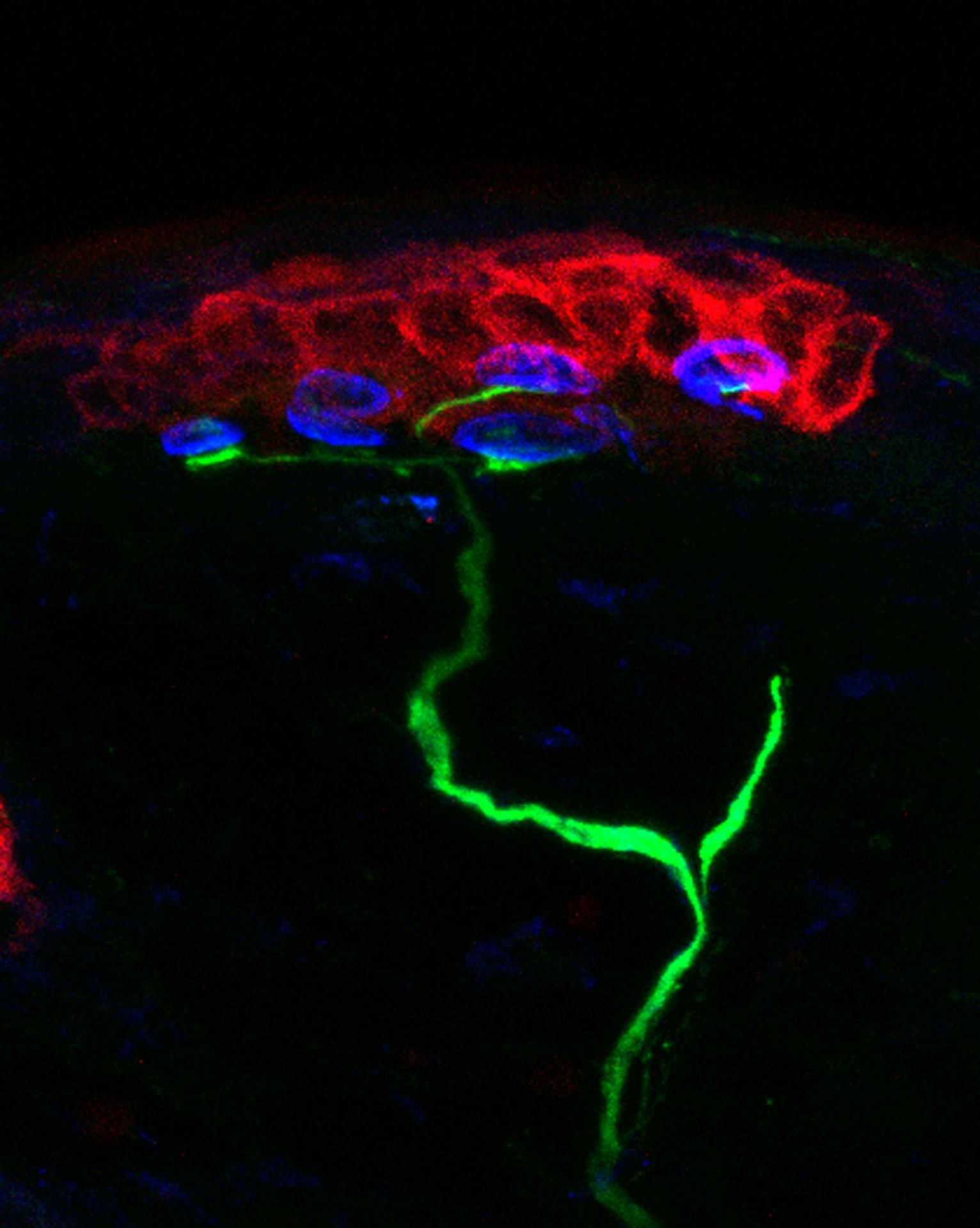
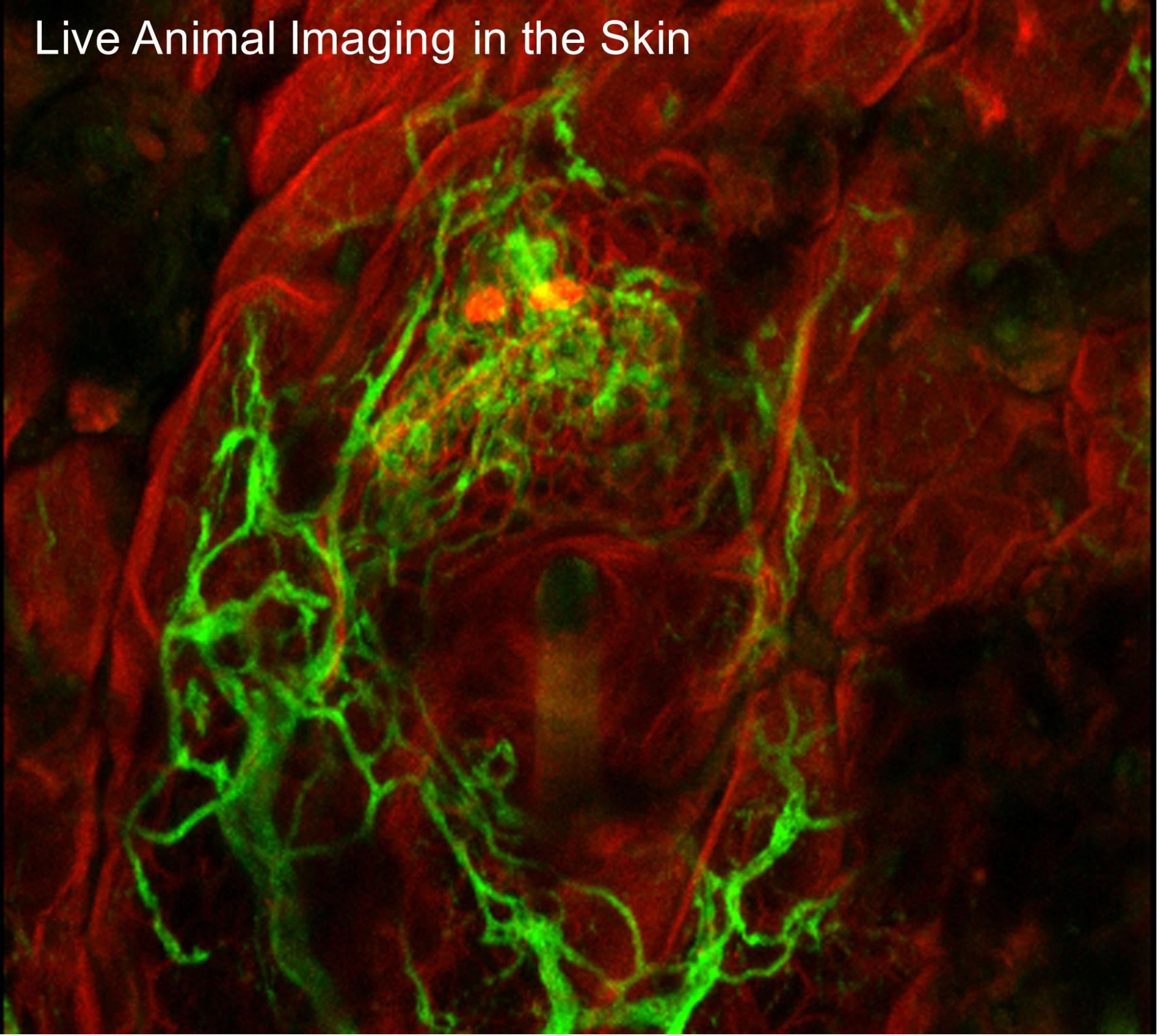
The SCIM Core provides state-of-the-art techniques for i) isolation, propagation and functional assessment of skin stem cell lineages, ii) induced pluripotent stem cell systems, iii) in utero gene targeting, iv) tissue culture systems to analyze genes related to skin diseases by efficient and systematic overexpression or suppression including CRISPR/Cas9-mediated deletion and v) three-dimensional imaging platforms specialized for skin.

The SCIM is committed to offering assistance to epiCURE investigators (experimental design/individual protocol development, interpretation of obtained results, teaching) and assuring effective communication among investigators in order to reduce the research costs, increase efficiency and further enrich the scientific quality of epiCURE.

The SCIM was specifically designed as resource to support a translational-based pipeline to facilitate skin disease studies that is able to bridge basic science with translational models. As such, SCIM services will be tailored to the needs of each investigator in a manner that is highly responsive to the Precision Medicine directive and the NIAMS mission.

The scope of services provided by the SCIM is outlined in the following Service Clusters:

Service Cluster A. Tissue Analysis

  • A1. Harvesting, processing, and phenotyping rodent and human skin specimens
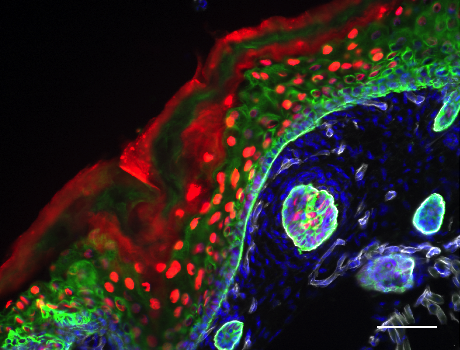
  • A2. Skin morphology, special histochemical and immunochemical analyses
  • A3. Accessing normal and lesional skin tissue banks
  • A4. Accessing neonatal human foreskin specimens


Service Cluster B. Cellular and Tissue Engineering

  • B1. Isolation and propagation of primary human or murine skin cell cultures
  • B2. Preparation of 3T3 fibroblast feeder cells
  • B3. Lentiviral and CRISPR/Cas9 engineered mouse and human skin cells
  • B4. In Utero Gene Targeting using RNAi gene knock out technology


Service Cluster C. Skin Stem Cell Analysis

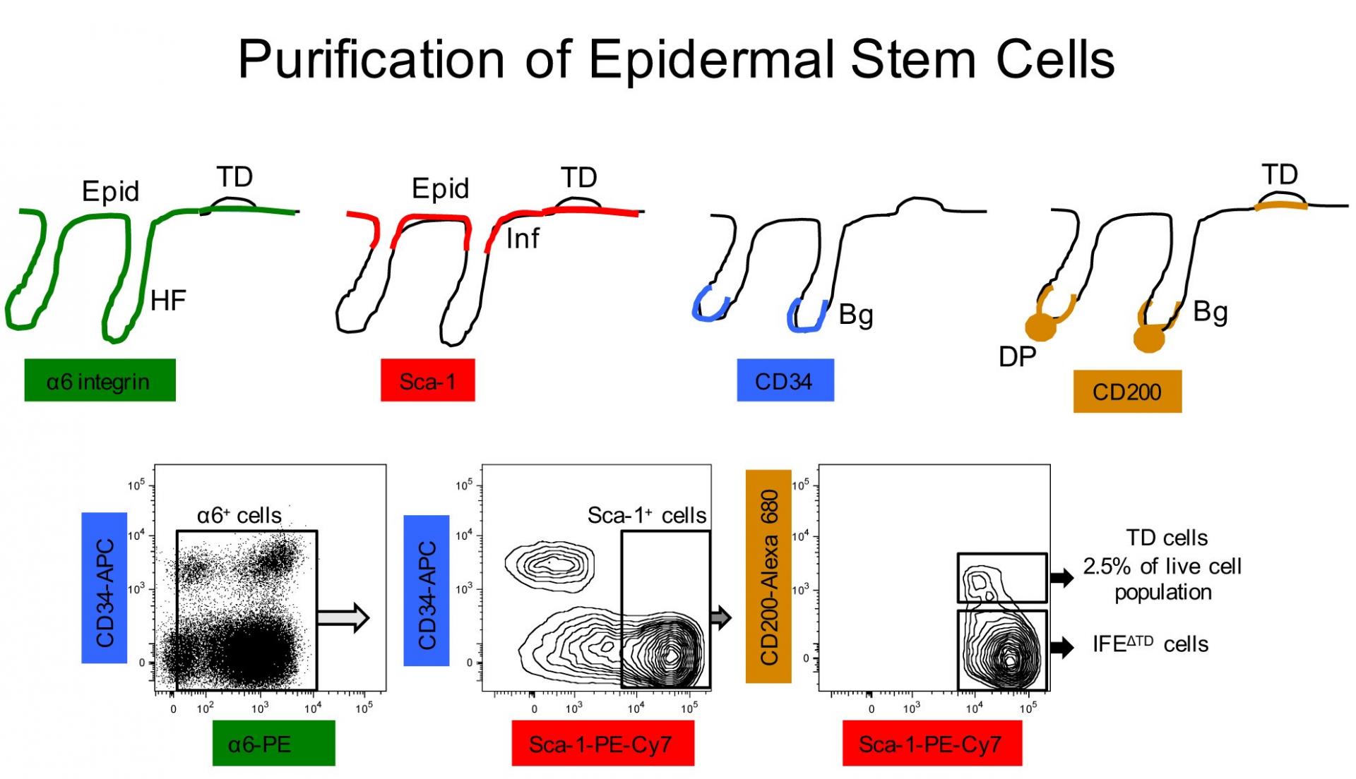
  • C1. Live stem cell isolation and in vitro clonogenic and proliferation (EdU) assays
  • C2. Hair and skin reconstitution assay using grafting chambers
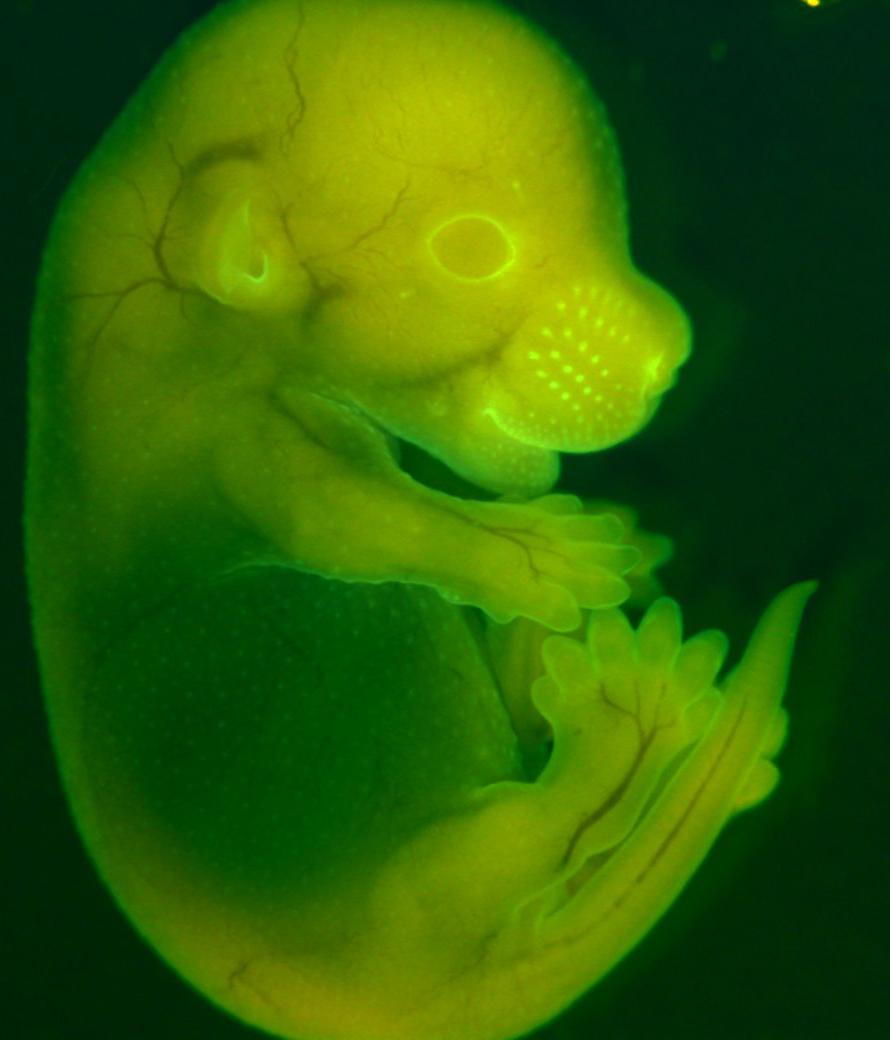
  • C3. In vivo lineage tracing/fate mapping and epidermal proliferation (EdU) analysis
  • C4. Propagation of iPSC derivatives from human skin fibroblasts or keratinocytes


Service Cluster D. Specialized In Vivo Imaging Platforms

  • D1. Three-dimensional structural imaging in skin whole mounts
  • D2. High-resolution cellular imaging in skin whole mounts
  • D3. Live animal skin multi-photon microscopy
  • D4. Skin Pathology Scoring and Tutorials